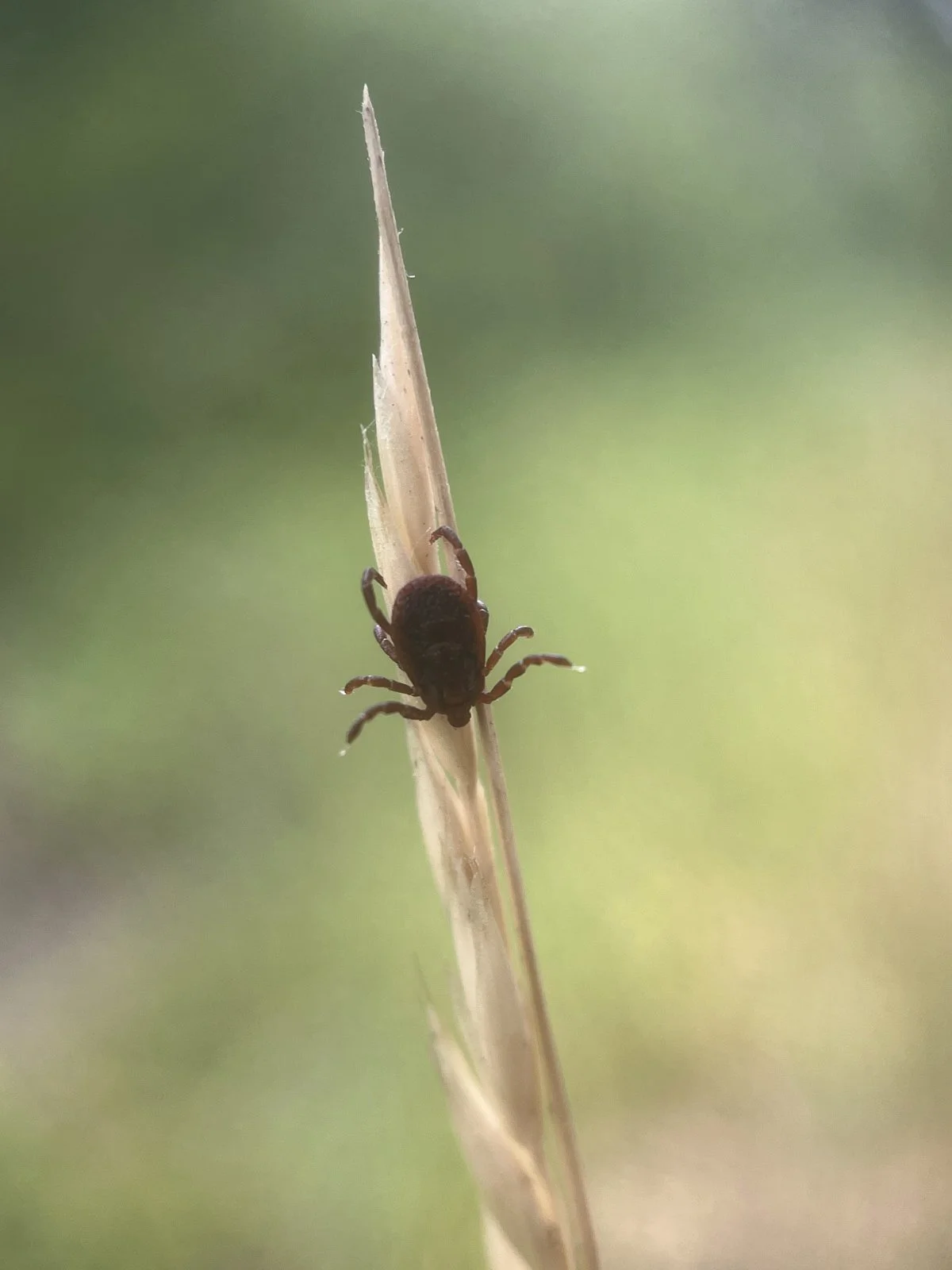
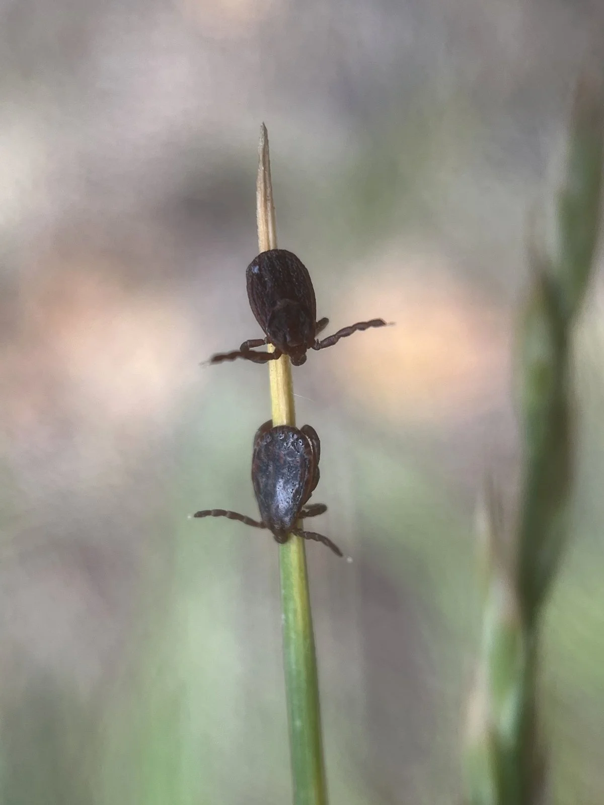
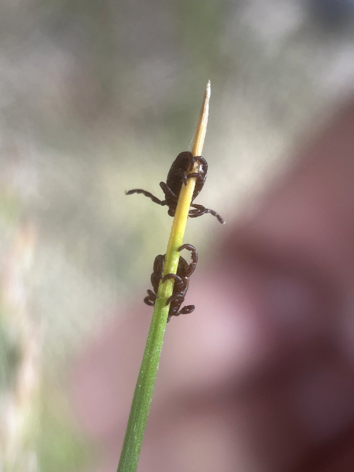
Mistletoe

Mistletoe can be seen growing in the Oregon White Oak trees out at the arboretum. They are evergreen and keep their fruit into the winter which is a beneficial food source for birds like Western Bluebirds.

Mistletoe is a hemiparasitic plant. This means that it takes water and nutrients from its oak tree host, but also contains chlorophyll and is able to perform photosynthesis to produce some of its own food. The amount that it performs photosynthesis varies once the plant becomes established and varies between the different species.


An oak tree recently fell at the arboretum next to the old barn. At the top of the tree there was some mistletoe growing which was now easily accessible as the tree lay on the ground. I trimmed off a small branch that had some mistletoe growing from it and took it over to the wood shop where Patrick neatly cut the mistletoe in half with a band saw so I could see what it looked like on the inside. It was really interesting and beautiful to see the internal structure of where the two plants grew together. Thanks Patrick!


Patrick removed the branches of the mistletoe so that it would fit through the saw. In the photo, you can see me holding the branch in my hand and the mistletoe growing out of the top. He made two vertical cuts, one through the mistletoe and one about an inch to the right on the oak branch.


In the photo, I am holding the mistletoe stem and showing you the part where the mistletoe and the branch were cut in half vertically. The top half is the mistletoe and the bottom half is the oak branch. When the mistletoe germinates, it grows down into the vascular tissue where it taps into the xylem and/or phloem. In general, most hemiparasites are primarily parasites of the xylem in order to obtain water and inorganic nutrients. Holoparasitic plants do not photosynthesize and therefore will tap into the phloem as well to obtain nutrients like sugars produced during photosynthesis from the leaves of the host plant.

It is interesting to see the woody tissue of the mistletoe and the oak branch growing together. Once the mistletoe establishes itself on the branch by growing rootlike structures in the vascular tissue, the mistletoe’s growth becomes synchronized with the growth of the oak branch. This coordinated growth area is called the meristem. Essentially, the mistletoe starts to grow like a “branch” on the oak tree limb. In the second photo, you can see that it has a woody stem, a thin layer of bark, and lichens start growing on it. Reading about the biology of the interaction between mistletoe and its host plant can be a bit complex, for me anyway, but that is the overall idea of it from what I understand.


There’s more! There’s also a structure growing beneath the bark of the oak limb growing longitudinally from the mistletoe. These are called cortical strands and help to further establish the mistletoe by gathering nutrients and anchoring it onto the branch. The cortical strands will start to send sinkers down into the branch as well to further assist in securing the mistletoe to the host. This root-like structure of cortical strands and sinkers comprise what is known as the endophytic system of the mistletoe.

Again, reading about all of the biology of the interaction between mistletoe and its host plant is complex, but this is my basic understanding of it. I recommend that you check it out and do some further research, maybe you’ll be saying to yourself, “C’mon Bryan. It’s not that complicated.”

My sincere hope is that this blog inspires you to go outside, check it out, and maybe do some more research into the topic. Nature is an inexhaustible source of wonder. I hope to see you out there!

Resources

Ehrenberg, Rachel. “Marvelous Misunderstood Mistletoe.” Knowable Magazine, 18 Dec. 2020, https://knowablemagazine.org/content/article/living-world/2020/marvelous-misunderstood-mistletoe.

Glatzel, G., and B. W. Geils. “Mistletoe Ecophysiology: Host–Parasite Interactions.” Botany, vol. 87, no. 1, Jan. 2009, pp. 10–15, https://doi.org/10.1139/b08-096.

Whiteman, Noah K. “Mistletoes.” Current Biology, vol. 33, no. 11, June 2023, pp. R467–69. ScienceDirect, https://doi.org/10.1016/j.cub.2023.03.035.

Springtail Takes a Bubble Bath

The other day I soaked some moss in a small dish for about 20 minutes to really hydrate the moss and energize a very tiny creature that lives in the moss called a waterbear. After soaking, I lightly stirred and shook the moss out in the dish and placed it under a microscope. While inspecting the dish for waterbears, I came across a springtail on the surface of the water that had been washed out of the moss into the dish. While watching it, I noticed that it was moving around on the top of the water tilting back and forth. I saw a bubble appear at its mouth and it retrieved it with one of its legs. It then proceeded to rub the bubble on its antennae and its head. It continued to produce more bubbles and groom its entire body. Occasionally it would attempt to spring away, but landed back into the dish. I would reposition it under the microscope and before too long it would start grooming again. Fascinating!

Springtails live in the moist environment created by the layer of leaves I spoke about in the previous post. Upon a little research, I found that they have a fluid bubble that they bathe with to keep themselves clean, especially their antennae which are their sensory organs. The fluid bubble is also necessary to waterproof themselves to help keep from drying out.

The springtail’s scientific name Collembola refers to their two inflatable tubes known as Collophores located on the underside of the first segment of the abdomen. They help in bathing by reaching the hard to reach places. They can also stick it to the ground to flip themselves over if they end up on their back after springing through the air. I found a small segment of a BBC documentary series called Life in the Undergrowth narrated by David Attenborough that talked about springtails. Click here to watch. It’s about two and half minutes long. This part about springtails was great, and I am looking forward to watching the whole series.

Springtails are important beings to a healthy ecosystem and have such interesting lives. They live right under our feet, but mostly go unseen and are out of our awareness. There can be up to 10,000 springtails in one cubic foot! I would love to see the environment where that many springtails are living so densely populated.

Nature is an inexhaustible source of wonder. I hope to see you out there!

Sources
“BBC One - Life in the Undergrowth, Invasion of the Land, Little Leapers.” BBC, 22 June 2009, https://www.bbc.co.uk/programmes/p003km37.

“Springtails.” Texas A&M AgriLife Extension Service, https://agrilifeextension.tamu.edu/library/gardening/springtails/. Accessed 13 Jan. 2026.

Today, Entomology. “The Collophore Helps Put the Spring in Springtails.” Entomology Today, 27 Aug. 2015, http://entomologytoday.org/2015/08/27/the-collophore-helps-put-the-spring-in-springtails/.

Spring in Winter

The winter sky is low. I feel like I have to duck under the shallow ceiling of amorphous, gray clouds as I walk around the arboretum. If the sun peaks through, it is just above the brim of my hat. In the hibernal landscape, I found an amazing “spring” hiding under a bed to put some bounce in your step to help alleviate any winter blues.

Beneath the oak trees, a patchwork of brown leaves blanketing the ground creates the perfect home for springtails! Oak leaves are high in tannins and lignin which causes them to decay at a slower rate. It can often take years for them to break down. This insulative bed of leaves helps regulate moisture and temperature creating a favorable home for many creatures in the leaves and the ground below. These leaves also add a layer of protection from potential predators by giving them a place to hide.

The other day I knelt down at the edge of the leaves and peeled back the top layer. I was immediately greeted by a lively burst of springtails emanating forth in every direction. A unique characteristic to most springtails is a forked tail-like structure called a furcula. It is folded underneath their abdomen and held in place by a latch called the tenaculum. When the tenaculum is released, the furcula quickly flips open. This acts as a spring as it pushes downward against a surface, launching the springtail into the air to hopefully evade predators.

Springtails are important detritivores. They primarily feast on decaying organic matter which cycles nutrients back into the ecosystem. In addition, as they burrow and wiggle around in the soil eating and searching for food, their small tunnels help build the soil microstructure. Along with decaying organic matter, they eat fungi, algae, and bacteria.

Springtails are tiny arthropods that until recently were considered insects. Around the year 2000, they were reclassified in the class Collembola. They are similar to insects but along with DNA testing, they have characteristics such as internal mouth parts and unique structures like a tube-like collophore and the aforementioned furcula that play a role in separating them out into the class Collembola.

They are one of the most abundant arthropods in the soil mesofauna and one of the most interesting. The other day I found what appears to be three different kinds in and under the oak leaves out at the arboretum. I am going to continue researching and exploring these wonderful creatures and I hope this small introduction inspires you to do the same.

Happy New Year!

Sources
“All about Springtails (Collembola).” A Chaos of Delight, https://www.chaosofdelight.org/collembola-springtails. Accessed 7 Jan. 2026.

Class Collembola – ENT 425 – General Entomology. https://genent.cals.ncsu.edu/insect-identification/class-collembola/. Accessed 7 Jan. 2026.

“Springtails.” Missouri Department of Conservation, https://mdc.mo.gov/discover-nature/field-guide/springtails. Accessed 7 Jan. 2026.

Springtails | Colorado State University Extension Website. 20 June 2025, https://extension.colostate.edu/resource/springtails/.

Western Meadowlark

There is a large meadow on the east side of Mt. Pisgah with a small creek running through it that carries rainwater off the hillside down to the Coast Fork of the Willamette river. Thickets of buckbrush fill in the slope as it rises out of the meadow with oaks scattered around the hillside. These layers create good habitat for a diversity of birds. One that I found over there this fall was the Western Meadowlark, the state bird of Oregon.

As I walked along the trail, I flushed a few hidden in the tall grass. Some flew up into to the lower limbs of nearby oak trees. From their perch in the tree, I could easily see their bright, yellow breast with a bold, dark ‘V.’ Some of them settled back down further up the hillside. As they flew over the meadow, their flight consisted of alternating short, rapid wing beats with glides as they flew just above the top of the grass. As they descended back onto the ground, their tail fanned out revealing a flash of white from their outer tail feathers. The upper side of their bodies are a mottled pattern of brown, black and buff, which allows them to easily disappear into the meadow.

As you may have noticed, meadowlarks have a pointed, stout bill that aids them in a foraging method called gaping. It inserts its bill into thick grass or the ground and then open its bill. This pries apart the grass or ground to reveal seeds or insects hidden underneath.

Western Meadowlarks are messengers of beauty and wonder. They have a melodious, warbling song that embodies the spirit of the West—vast landscapes and open skies. I hope you have had the opportunity to hear one sing.

On its website, The Cornell Lab of Ornithology had this fact that I thought you might include in some cocktail party conversation during your holiday celebrations: “John James Audubon gave the Western Meadowlark its scientific name, Sturnella (starling-like) neglecta, claiming that most explorers and settlers who ventured west of the Mississippi after Lewis and Clark had overlooked this common bird.” Honestly, I think Audubon flubbed this one. This bird’s Latin name could be changed to something that appropriately reflects its spirit and beauty.

Happy Solstice

Happy Solstice! I hope that you find time to be out in nature. I wish you peace and good health in your body, mind, and spirit.

Nebulous Tangerine Dream

While researching biosheens for the last post, I came across iron bacteria as a possible organism that creates them. When I looked them up, I saw that they produce a brilliant orange cloud in the water which I didn’t see around the bridge where I witnessed the biosheen. So I went back out to the arboretum to see if I could find this nebulous, tangerine dream.

Iron is the fourth most common element in the earth’s crust and vital to life. The biogeochemical cycle of iron through the biosphere, atmosphere, lithosphere, and hydrosphere is often cleverly referred to as the Ferrous Wheel. Iron is constantly cycling through the environment in two states, ferrous iron (Fe2+) and ferric iron (Fe3+). Ferrous iron has a positive charge of plus two because it has lost two electrons and ferric iron has a positive charge of three.

This orange cloud is a result of oxidation (losing electrons) by iron bacteria converting ferrous iron (Fe2+) into ferric iron (Fe3+). The most common acceptor of this electron is oxygen. Ferrous iron is highly dissolvable in water in anerobic (oxygen-free) environments. Where this water leaches out into aerobic (oxygen-rich) environments, iron bacteria thrives. Iron bacteria obtains energy when it transfers an electron from ferrous iron to oxygen creating ferric iron which is insoluble in water. This insolubility creates a beautiful orange soup of ferric iron and iron bacteria swirling around in the water.

For me, the ferrous wheel was a fascinating and dizzying ride full of science that I spent lots of time reading about. The importance of iron will surely find its way into more posts in the future. For example, iron is vital for the production of chlorophyll which is a key component for photosynthesis. For now, I feel this amount of information is enough to get you started doing some research of your own, good luck.

Also part of this story, I put my hand down in the water to see how it felt. It looked slimy, but I was unable to feel it. I expected it to cling to my hand when I pulled it out, but it didn’t stick to my hand at all. I scooped up some in a container to have a look. I immediately noticed that I had caught a small organism and it was excitedly darting around. That creature was me swimming around in this nebulous, tangerine dream—it was a metaphor, a reminder of a world so unbelievable in its infinite complexity and bewilderment.

Nature is an inexhaustible source of wonder. I hope to see you out there!

Biosheen on Wetlands

During the first couple of weeks of November, the surface of the water was covered with a thin film that had become fractured and had a colorful sheen suggestive of an oily substance.

One way biological surface sheens can occur is the result of bacteria decomposing organic matter. Places like the wetlands become a suitable environment for bacteria to flourish because the flow of water is very low or not at all which can lead to low levels of oxygen in the water. With the heaps of newly added leaves from deciduous trees and shrubs growing in the wetlands, the perfect, soupy environment has been created to break down all this plant material and create a biosheen.

A biological sheen can appear similar to a sheen caused by petroleum. To tell the difference, a biological sheen breaks into irregular or jagged edges when the surface of the water is disturbed and petroleum sheens stays together in a slick.

After I made time to do a little research on the phenomenon, I went back to area to investigate if it felt oily, had an odor, etc. Disappointingly, I found that it had completely disappeared. I stood there wondering about it and wished I had been more curious earlier. Where did it go? Why was its presence so brief? Did it dissovle? Did another organism eat it?

I have been looking around the internet and haven’t come across more specifics on how biosheens are created as a byproduct of decomposition and more details of their makeup. I will leave it there at that moment as I continue to solve this puzzle.

Nature is an inexhaustible source of wonder. I look forward to seeing you out there.

Coyote Scat - Grapes?

On the road through the meadow, I came across some coyote scat that blended in well with gravel surface. I was a little disappointed to see that they changed their diet, and I wasn’t seeing any more of the bright orange apple scat. The menu appears to have switched over as the apples ran out, now it appears that they are eating grapes. There is a vineyard on the other side of the river from the arboretum. The water is still pretty shallow and the current isn’t very swift so they could easily cross, or they could always use the bridge.

Coyotes are opportunistic predators that eat a wide variety of foods that includes lots of fruits. When I found the apple scat, it was interesting to see sizable chunks of apple fairly undigested. In this pile of scat, I found a berry that had been swallowed, passed through the coyote’s digestion system and out the other side as a whole grape! I rolled it out of the scat and realized that somehow it hadn’t been smashed or punctured, which doesn’t seem possible. In one of the photos below, I rolled the grape over to the side and put a couple of seeds beside it. The arboretum caretakers Patrick and Conner came by as I was investigating the scat and they thought it looked like grapes in the scat too. Patrick thought I should go over to the vineyard and see what kind of grapes are growing and compare them. That’s a good idea. Afterwards, I gently pushed on the berry with a stick. It popped and the juice oozed out with a couple of seeds. I wondered what surprises might show up next in the coyote scat. Do coyotes eat rosehips?

Pholiota Mushroom

This cluster of golden mushrooms vibrantly glowed in the afternoon sun this fall. They were popping out of the side of a willow tree growing in the wetlands. Its sunny, yellow-orange cap was adorned with orange scales and was moist and sticky. This gelatinous quality of the cap was referred to as viscid in the mushroom guides I have. Underneath, it had wonderful gills radiating out from a scaly stalk.

This mushroom is classified as a saprobe—a group of fungi that act as decomposers. They get their nutrients from feeding on dead and decaying wood, leaves, litter, and other organic matter. It was interesting to see them growing from the sturdy trunk of a live tree. I wondered if a mischievous forest gnome had glued the stalks to the side of the tree. Later, I read that some of the species of Pholiota grow on live trees. That said, farther up the tree on the end, it was damaged and decaying which maybe has worked its way down into the center of the tree where these mushrooms were emanating.

Identifying mushrooms is tricky business. Every time I read about them, the guide books refer to look-alikes and needing to look at spores along with other factors such as habitat, time of year, etc. So I only feel comfortable identifying this one down to the genus Pholiota.

I have been struck by the abundance and variety of mushrooms this year. I wondered if the weather conditions this year had brought forth more mushrooms. This seemingly would make it easier to notice ones that are usually there this time of year because they are in greater numbers. Either way, I am especially enjoying encountering them throughout the landscape this fall.

Fungus Gnats

A little flurry of insects whirled upwards when I approached this mushroom to have a closer look. I sat down for a few moments and they settled back down. I had heard about fungus gnats before and I wondered if these tiny insects on this mushroom could be them. As I attempted to take a few photos, I realized that they weren’t all the same. Maybe I found a tiny mixed flock of gnats just like small songbirds form in the winter. As I sit here at my desk trying to discover what these tiny creatures are, I realized that identifying small insects is challenging. So for now, I have decided to let them flit around in my curiosity and let them settle in the not sure category.

That said, I will give you a little information on what I learned about fungus gnats. They are decomposers and help to recycle nutrients back into the ecosystem. In their larval stage, they consume things such as fungi, algae, root hairs, and decaying plant matter in the soil. The adult fungus gnats eat very little, only consuming water or plant nectar. The adults are drawn to mushrooms because of the ideal, damp habitat for reproduction. It provides for a suitable place to lay their eggs and have the larvae hatch in a moist environment rich in organic material in the soil to feed upon. Fungus gnats also provide a food source for animals such as birds like all of the kinglets here at the arboretum for the winter.

Interestingly, when investing many insects, much of the information out there on the internet begins with defining them based on if they are “harmful” or “beneficial.” In other words, do they “damage” plants that people are trying to grow for food, clothes, housing, such as, apples, cotton, or trees, or are they “helpful” in eating these “harmful” insects. I read that fungus gnats can infest greenhouses or mushroom operations but I didn’t see it mentioned happening out in nature.

We need to start shifting back to the paradigm of living in harmony with the earth where all life is respected. Where we are filled with thanksgiving, wonder, curiosity, and awe at all of the beauty and diversity of life. Where we see ourselves as a part of nature and its caretakers for future generations. Thank you to all the people out with there trying to make it happen. Thank you for reading my blog.

Nature is an inexhaustible source of wonder. I hope to see you out there!

Hare's Foot Inkcap

There have been heaps of these mushrooms growing out of a blanket of wood chips that was scattered on the ground behind the White Oak Pavilion during the cleanup process after the ice storm a couple of years ago. I am really new at trying to identify mushrooms and many can only be reliably separated with microscopic examination. I think these are hare’s foot inky caps, Coprinopsis lagopus, because they are a common mushroom known for appearing in urban settings on wood chips and they fit the description and match the photos in the field guide. As it first emerges, it looks like a hare’s foot, but it quickly loses that fuzzy resemblance. I generally saw them last only for about a day or less as they quickly mature and turn into a black, ink-like mass.

In Mushrooms Demystified David Arora says this about inky cap mushrooms:
”MEMBERS of this genus are called inky caps because the gills and often the cap digest themselves at maturity, turning into an inky black fluid that drips to the ground. The autodigestion or deliquescing of the gills plus the black spore print are the main diagnostic features of Coprinus… The autodigestion process is a unique method of spore dispersal that should not be confused with the normal process of decay that occurs in most mushrooms. Rather than maturing at an even rate, the spores near the margin of the cap ripen first. Enzymes are simultaneously released which dissolve the surrounding tissue, causing the edge of the cap to spread out and curl back. This pulls the gills apart, enabling the spores to be discharged into the air…”

The edition of Mushrooms Demystified that I quoted is from 1986. Due to recent DNA studies, the genus of mushrooms Coprinus has been split into four genera: Coprinus, Parasola, Coprinopsis, and Coprinellus. At the time I guess it made sense to group of all these mushrooms together based on this characteristic process of spore dispersal as the mushrooms matured and turned into an inky substance.

Inky caps are saprotrophs which are organisms that derive nutrients by decomposing dead or decaying organic matter. They are essential for a healthy ecosystem in recycling nutrients back into the soil for other organisms to use.

I loved seeing all of the beautiful forms that this mushroom underwent as it quickly grew and matured. Check out all of the photos I took below over a couple weeks of returning to this spot over and over.

Resources
Arora, David. Mushrooms Demystified: A Comprehensive Guide to the Fleshy Fungi. Second edition, Ten Speed Press, 1986. K10plus ISBN.

Coprinoid Mushrooms: The Inky Caps (MushroomExpert.Com). https://www.mushroomexpert.com/coprinoid.html. Accessed 19 Nov. 2025.

Trudell, Steve, et al. Mushrooms of the Pacific Northwest. Timber Press, 2009. Timber Press Field Guide. Open WorldCat.

Fruiting Fertile Earth

The essence of the earth is flowing out of the center of this mushroom cap with its rich, fertile brown. A fruiting body conjured up by the spirit of the rain—a reminder of a wondrous, subterranean world. Small creatures gathered under its umbrella. Had they come to mingle and dine, to give thanks to the mystery of life and to honor Mother Nature’s wellspring of beauty?

California Poppy Fungus

Upon seeing the orange peel fungus, a friend of mine thought that it looked like a California poppy flower blooming out of the ground. I agree. Lots of organisms in nature have multiple common names, and I think California poppy fungus would be an apt name to add to the list. They are both in bloom at the moment at the arboretum and have a beautiful orange glow in the afternoon sun.

Thimbleberry Gall Wasp

While out walking in the arboretum the other day, I got caught in a little eddy of flitting and chirping. I was on the pond lily trail and a mixed flock of birds was excitedly gobbling down woolly aphids. There were chickadees, ruby-crowned and golden-crowned kinglets, brown creepers and Bewick’s wrens. As I turned to watch a ruby-crowned kinglet bouncing between the branches near the ground, I noticed a little knobby knuckle on a small stem. It was firm and woody and had small holes on its surface as if an insect had chewed its way out. As I looked closer at the surrounding vegetation, I saw a few more and realized that they were on thimbleberry stems. I looked them up to find that they were thimbleberry galls created by the wasp Diastrophus kincaidii.

I found an informative paper online written by James K. Wangberg called “Biology of the Tliimbleberry Gallmaker Diastrophus kincaidii.” It was published in 1975 in the journal The Pan-Pacific Entomologist. When writing about the life cycle of D. kincaidi, he writes that the adults emerge from the gall in the spring by chewing their way out. I cut one open to see what it looked like inside and found that it still had tiny larvae inside. When I saw this gall riddled with holes, I thought that the wasps had already left. I thought that perhaps that some of the larvae were still developing and hadn’t emerged from the gall yet. I hadn’t read Wangberg’s study yet, so I didn’t know that the adults chew their way out in the spring. So why did these galls have holes in them?

At the end of Wangberg’s article he had a section called “Insect Associates,” which states: “Ten species of parasitic hymenopterans were found associated with D. kincaidii on thimbleberry. Most are parasites of D. kincaidii but some are hyperparasites. Parasitism of D. kincaidii larvae is extensive during the summer and oftentimes only those individuals deepest within the gall tissue escape attack. The parasitic species emerge in succession throughout the summer and D. kincaidii is subject to parasitism for its entire larval life. In addition, an inquilinous weevil and an undetermined cecidomyiid midge were occasionally present within galls.”

Its a wonder that any of the original gall wasp D. kincaidii survive to adulthood with that many parasites. So were the holes created by the parasites piercing the gall and laying eggs or was it from them chewing their way out? My guess is that it was from chewing their way out. The gall is created on new, green stems and becomes woody as the season progresses into fall. I imagine the parasitic wasps pierced the gall when it was softer and green and the nice round hole was neatly made during exiting. So, are the larvae that are still inside the gall I cut open surviving D. kincaidii or one of the parasites?

I’ll leave you with a quote by Ken Kesey that is on a memorial stone at the arboretum: “The answer is never the answer. What's really interesting is the mystery. If you seek the mystery instead of the answer, you'll always be seeking. I've never seen anybody really find the answer. They think they have, so they stop thinking. But the job is to seek mystery, evoke mystery, plant a garden in which strange plants grow and mysteries bloom. The need for mystery is greater than the need for an answer.”

Vocabulary
inquiline: an animal exploiting the living space of another, e.g. an insect that lays its eggs in a gall produced by another.

Resource
Wangberg, James K. “Biology of the Tliimbleberry Gallmaker Diastrophus Kincaidii.” The Pan-Pacific Entomologist, vol. 51, no. 1, Jan. 1975, pp. 39–48.
Link to article

Diversity & Beauty of Fungus

The diversity and beauty of fungus is miraculous. While walking on the hillside trail looking for Douglas-fir cone fungus Strobilurus trullisatus (Post: Douglas-fir Cone Fungus, 11/25/24), I came across this sparkling Douglas-fir cone. I knelt down on the ground to take a closer look. I magnified it with my camera and found that it was frosted with small, white cups that appeared to have tiny beads of water clinging to their surfaces. Their translucent, white bodies glowed against the dark brown scales of the cone. A fungus had found a home inside of this Douglas-fir cone and blossomed during the recent rains. It decorated its humble abode with small fairy lights to brighten our paths in the waning light and pale gray skies of fall in the Northwest. Thank you.

Flies: Syrphidae Family

Over the past couple of weeks I have been seeing tons of woolly aphids floating through the air. So I decided to follow them around to see where they were going and take a photo of one when it landed. I found some of them landing on ash branches and snapped a couple of photos.

Wait, let me back up a little. The reason I was chasing down aphids was because I came across this black and yellow fly with dark red eyes hovering around. I thought they were beautiful as they perched on vegetation in the sunlight. I was lucky enough to get a photo of one and I put the photo in iNaturalist. A list of a bunch of flies came up in the Syrphidae family. These are commonly called syrphids or hover flies. Some of the identification suggestions listed for my photo included Common Loopwing Aphideater, Western Aphideater, Black-tailed Aphideater and Bare-winged Aphideater. iNaturalist listed 10 suggestions and they all were black and yellow syrphid flies that looked very similar to me. So, I thought I would do a little reading on syrphid flies in general. I discovered that the primary diet of the larval stage of many syrphids is aphids!

The adults are not predators. Their diet mainly consists of flower nectar and pollen, consequently they are important pollinators. Finding them on flowers combined with their size and coloration, they can be mistaken for bees and wasps. Syrphid flies don’t sting or bite so this mimicry is a form of protection from predators. Also, the diet of adult syrphids can include the sugary honeydew secreted by aphids.

So, back to the beginning of this post. I followed the aphids around to see where they were going and thought I might find some syrphid flies hovering around. I did! Being that aphids are the syrphid larvae’s primary food source, they want to lay their eggs near aphids.

Syrphid flies are quick fliers and I was unable to follow them for very long as they zoomed through the landscape. So I wasn’t able to see them laying eggs. I also realized that this endeavor is probably best pursued during the warmer days of late spring and summer when I see more aphid colonies congregating on leaves and probably more syrphids nectaring nearby on flowers. Some species of syrphids overwinter as adults, but most overwinter in the larval or pupal stage in protected areas such as leaf litter. In moderate climates, they can be seen year round.

So next year I will be looking more closely and hopefully I will see the syrphids laying eggs and larvae eating aphids!

Nature is an inexhaustible source of wonder. I hope to see you out there!

Resources
Syrphid Flies. https://extension.umn.edu/beneficial-insects/syrphid-flies. Accessed 5 Nov. 2025.

Syrphid Flies | NC State Extension Publications. https://content.ces.ncsu.edu/syrphid-flies. Accessed 5 Nov. 2025.

Syrphids (Flower Flies, or Hover Flies) / Home and Landscape / UC Statewide IPM Program (UC IPM). https://ipm.ucanr.edu/natural-enemies/syrphids/#gsc.tab=0. Accessed 5 Nov. 2025.

Flicker Feathers

While picking up trash by the river, I came across a pile of flicker feathers under a small thicket of willows. It was a mix of emotions to find them—saddened to see that a beautiful bird had been killed and fascinated at the beauty and patterns of all the feathers. It was also a mystery. There were so many feathers. It was like the predator took the time to neatly pluck all of the feathers out before it ate the flicker. There wasn’t any sign of the body of the bird and there wasn’t any blood on the ground or on any of the feathers. It felt like the predator recognized the beauty of the bird and wanted to honor the life it had taken by carefully plucking the feathers to preserve their beauty.

Candle-snuff Fungus

The fruiting bodies of fungus have an unbelievable amount of diversity. With its charred, slender body and ashen-white tip, Xylaria hypoxylon is likened to the wick of a snuffed out candle.

In Mushrooms Demystified David Arora describes the fruiting body as “2-8 cm high, very tough, erect, slender, cylindrical or narrowly club-like but usually becoming anterlike (branched sparsely or forked at the tip) in age.” Its body did feel very stiff and tough to the touch which was different than what I expected. I tend to think of mushrooms as being mostly tender, so I was a little surprised by how sturdy and sinewy this one felt. The powdery white branch tips are the asexual spores and contrast beautifully with the black stalk.

David Arora describes its habitat as: “Scattered to densely gregarious or clustered on rotting logs, stumps, buried sticks, etc; widely distributed and common.” In this little patch, I found them on small sticks and on wood chips that were probably spread there during all of the chipping of branches after the ice storm a couple winters ago. This is a small mushroom and they blend in well. It took my eyes a little time to adjust to seeing them in all of the litter of fir needles, twigs, leaves, grass, and other vegetation on the ground.

Good luck on your quest to find them. I hope you are enjoying the fall.

Resources
Arora, David. Mushrooms Demystified: A Comprehensive Guide to the Fleshy Fungi. Second edition, Ten Speed Press, 1986.

Trudell, Steve, et al. Mushrooms of the Pacific Northwest. Timber Press, 2009. Timber Press Field Guide.

Coyote Scat - Apples?

Coyotes are opportunistic eaters with a diverse diet that includes small rodents such as mice and voles, rabbits, birds, carrion, deer, insects, lizards, fish, fruit, seeds and grass. This varied diet depends on the time of year and ecosystem that they are inhabiting.

Over the last couple of weeks out at the arboretum I have found coyote scat that consists mainly of what appears to be apples. When I broke some apart, it was full of what looks like apple skins, chunks of undigested apple pieces, and seeds. First and foremost, I can’t help being drawn over to the bright orange glow the coyotes are contributing to the fall colors. Why is it so beautifully orange?

Its scat is prominently displayed in places like in the middle of trails, trail junctions, on roadsides, or on rocks. These locations are chosen to communicate information to other coyotes, such as marking its territory. The last photo below shows a pile of scat at the edge of the gravel road where a path comes down from the meadow. I found multiple piles of feces in this area.

Curiously, it seems like the apples were only partially digested. That might be due to the coyote’s canine teeth chomping the apple into fairly big chunks as it ate and swallowed it. Perhaps, those bigger pieces just don’t get broken down as well as it passes through the coyotes digestive tract.

Nature is an inexhaustible source of wonder. I look forward to seeing you out there.

Powder-headed Tube Lichen

While walking down the path the other day, I came across a lichen that had fallen out of a tree. It looked like a small piece of coral from the ocean with its aquamarine color and tubular, branching lobes.

I picked it up to take a look and it was papery dry and light as a feather. It seemed quite delicate, and I felt I had to handle it with care to keep from crushing it. There was a small breeze and I slightly cradled it to keep it from blowing out of my hand. The tips of the tubular lobes develop into a nodule-like head called the soralia where the powdery soredia are produced, hence the common name powder-headed tube lichen. Soredia are asexual reproductive structures that contain both the photobiont (algae or cyanobacteria) and the mycobiont (fungus). The soredia will disperse and grow into a new lichen if they land in a suitable habitat.

Rhizines are fungal filaments that function to attach and hold many lichens to their substrate and don’t have vascular capabilities like the roots in plants. The Latin name of Powder-headed Tube Lichen is Hypogymnia tubulosa. Its genus is derived from the Greek words hupó, meaning “under” and gumnós, meaning “naked.” If you look on the underside of Hypogymnia tubulosa it has a blackened, wrinkled surface that lacks rhizines which is probably what this is referring to. So far in my research, I haven’t found a description of how its underside attaches itself to a substrate like tree bark.

I found a bare spot on a tree limb and nestled it between some other lichens to hold it in place. I wanted to see if it would reattach and continue to grow.

Nature is an inexhaustible source of wonder. I look forward to seeing you out there.

Sources
Hypogymnia.” Wikipedia, 22 Nov. 2024. Wikipedia, https://en.wikipedia.org/w/index.php?title=Hypogymnia&oldid=1259003117.

Jackson, August. “Lichens of Mount Pisgah: A Field Guide to Select Species.” Nov. 2022, https://mountpisgaharboretum.org/wp-content/uploads/Lichen-Field-Guide.pdf.

Lichen Biology. https://www.fs.usda.gov/wildflowers/beauty/lichens/biology.shtml. Accessed 21 Oct. 2025.